Transmission Electron Microscopy (TEM)
Info:cimemet@conicet.gov.ar

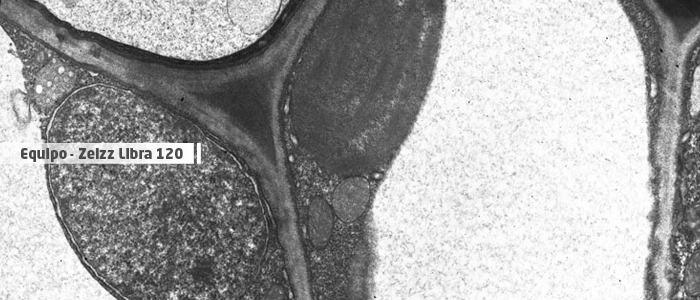
The Clear Field Transmission (BFTEM) images show the internal structure of the sample, and are obtained by selecting the electrons that cross it without deviating or with a minimum deviation. The contrast of the images obtained in this case depends on the thickness, density and atomic elements of the different areas observed, since we will obtain greater or lesser brightness depending on the less or greater difficulty that electrons have to cross the sample. The image obtained is similar to an x-ray of the sample, highlighting the inside of the sample as opposed to the scanning image (SEM) that reveals the surface.
TEM-DF In order to obtain Dark Field transmission images (DFTEM, Dark Field TEM), the electrons that are dispersed when passing through the sample are selected, blocking the direct beam (responsible for the Light Field) by opening the lens. In the image appear as bright areas those that disperse the electrons in the angle and direction that we have selected, appearing the dark rest. It is very useful for example to reveal crystalline particles within amorphous materials or to show defects in crystalline structures (dislocations, precipitates, etc.). Here you can see the previous image taken with the dark field technique, which reveals the existence of crystalline particles within the fibers.